
Photoacoustic imaging uses light and sound to 3D images and can be used to look inside the human body and provide clinical information.
Endoscopy is a test to look inside a body. The technology uses a long, thin tube with a camera inside called an endoscope. According to Dr Wenfeng Xia, research lead from the School of Biomedical Engineering & Imaging Sciences, “Traditional light-based endoscopes can only resolve tissue anatomical information on the surface and tend to have large footprints.”

Researchers have developed a way to exceed this limitation of the traditional endoscopy method. They have created a photoacoustic imaging endoscopy probe that can fit inside a needle with a diameter of 0.6mm. This device can resolve subcellular-scale tissue structural and molecular information in real time 3D images. This data technology allows a medical practitioner to identify and characterize the tissue during the procedure.
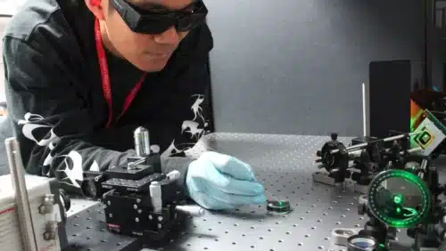
Photoacoustic imaging uses shining pulses of light with acoustic waves. The light is focused on the structures in the body and the ultrasound sensors detect the acoustic waves to form 3D images for structural and functional information for clinical purposes. Although this technology requires a bulky ultrasound detector and has low imaging speed, this issue has been resolved. Researchers combined wavefront-based beam shaping with light-based ultrasound detection and a fast algorithm for controlling the device. This unique combination allowed them to create an extremely small probe without sacrificing imaging speed.
The device consists of two optical fibers for delivering photoacoustic waves and another for detecting ultrasound waves. A high speed digital micro-mirror with nearly one million tiny mirrors is used for light to scan precisely.
To demonstrate the new device, the researchers used it to acquire high-resolution images of mouse red blood cells covering an area 100 microns in diameter. “We were able to accomplish this at about 3 frames per second,” Dr Xia said. “We also showed that the needle probe can be scanned to significantly enlarge the field-of-view in real-time by stitching together consecutive images.”
The technology has a broad range of applications, such as fluorescent imaging, Raman microscopy and two-photon microscopy. “It could eventually allow 3D characterization of tissue during various minimally invasive procedures such as tumor biopsies. This could help clinicians pinpoint the right area to sample, which would increase the diagnosis accuracy.”