

Researchers developed a deep learning algorithm to identify and outline lung cancer tumors for faster diagnosis and treatment
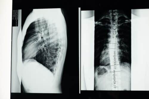
Researchers used an Artificial Intelligence algorithm to target problems caused due to conventional radiation therapy techniques, which makes the process easier, convenient, and faster. The validated deep learning algorithm traces and contours a non-small cell lung cancer (NSCLC) tumor on computed tomography (CT) scan within seconds. They used CT images of 787 patients to prepare their model to differentiate between tumors and other tissues. They assessed the performance of the scans on 1,300 patients. Further improvement in the algorithm needed the combined involvement of data scientists and radiation oncologists. Hence, whenever the researchers noticed any error in working with the algorithm suggested by radiation oncologists, they retrained the model using more scans to improve its performance.
“The biggest translation gap in AI applications to medicine is the failure to study how to use AI to improve human clinicians, and vice versa,” said corresponding author Raymond Mak, MD, of Brigham’s Department of Radiation Oncology. “We’re studying how to make human-AI partnerships and collaborations that result in better outcomes for patients. The benefits of this approach for patients include greater consistency in segmenting tumors and accelerated times to treatment. The clinician benefits include a reduction in mundane but difficult computer work, which can reduce burnout and increase the time they can spend with patients.”
Nowadays Lung cancer is most common and is treated using radiation therapy in almost one-half of cases. Radiation therapy is an ultra-long process that takes up to days to weeks for completion and also needs highly trained physicians to determine tissue target areas for radiation treatment. The researchers urged 8 radiation oncologists to carry out segmentation tasks and simultaneously rate and edit segmentation obtained by another expert or physician or algorithm. It proved that physicians could work 65% faster with 32% less variation using AI-produced segmentation compared to manually produced one, without compromising on performance. The quality of AI-drawn segmentation was better than human expert-drawn segmentation.
Now researchers plan to develop AI models that can identify “organs at risk” of receiving undesired radiation during cancer treatment and exclude them from radiotherapy. They ensure that AI-partnership helps clinical practice, by developing an independent segmentation algorithm that can differentiate between human and AI-drawn segmentation. “This study presents a novel evaluation strategy for AI models that emphasizes the importance of human-AI collaboration,” said co-author Hugo Aerts, Ph.D., of the Department of Radiation Oncology. “This is especially necessary because in silico (computer-modeled) evaluations can give different results than clinical evaluations. Our approach can help pave the way towards clinical deployment.”
Click here for the Published Research Paper