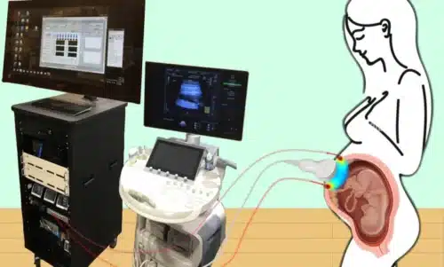
Researchers details a novel method for imaging the placenta in pregnancy and monitoring the oxygen levels of this crucial organ.

Researchers advanced the design of their instrument for almost a span of three years by consolidating the optical fibers and ultrasound probes into multimodal technology and exploring numerous ultrasound transducers to obtain stable and accurate measurements that also have reproducible features while obtaining clinical data. The design invented had capabilities to allow detailed study of the anatomy of the placenta and to obtain important details related to the functioning of the placenta such as blood flow, and oxygen levels which are difficult to acquire with current instruments.
The placenta is situated deep below the body surface and hence it is very difficult to get crucial information about the functioning of the placenta due to technical complications. These issues were addressed by Wang, a postdoc in Yodh’s lab, reducing the background noise in the optoelectronic system. Yodh says the key to success was to reduce background interference so that the small amount of light that penetrates deep into the placenta and then returns is still large enough for a high-quality measurement.
“We’re sending a light signal that goes through the same deep tissues as the ultrasound. The extremely small amount of light that returns to the surface probe is then used to accurately assess tissue properties, which is only possible with very stable lasers, optics, and detectors,” says Yodh. “Lin had to overcome many barriers to improve the signal-to-noise ratio to the point where we trusted our data.”
“Not only do we show that oxygen levels go up when you give the mom oxygen, but when we analyze the data, both for clinical outcomes and pathology, patients with maternal vascular malperfusion did not have as much of an increase in oxygen compared to patients with normal placentas,” says Schwartz. “What was exciting is that not only did we get an instrument to probe deeper than commercial devices, but we also obtained an early signal that hyperoxygenation experiments can differentiate a healthy placenta from a diseased placenta.”
The device is still under development and researchers are in process of refining the instrument to make it faster and user-friendly. As many clinical questions related to the placenta are unanswered, for Schwartz the biggest objective of this work is to furnish a way to get those answers. “Without being able to study the placenta directly, we are relying on very indirect science,” he says. “This is a tool that helps us study the underlying physiology of pregnancy so we can more strategically study interventions that can help support good pregnancy outcomes.”
Click here for the published Research Paper


