
The wearable device, created for bladder and kidney health monitoring, has the potential for early detection of deep-seated cancers in the body.
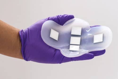
Photo courtesy of the researchers.
MIT researchers have created a wearable ultrasound patch that images internal organs without an operator or gel. It accurately images the bladder, aiding those with bladder or kidney issues. Adjustable in array location and frequency, it can also monitor other organs and potentially detect cancers like ovarian cancer earlier, improving diagnostics.

Wearable monitoring
The team has innovated a wearable ultrasound patch that sticks to the skin to image internal organs, with a focus on the bladder for their initial demonstration. This development mainly benefits patients with bladder or kidney conditions, offering a precise measure of bladder fullness. This patch is an alternative to traditional, bulky ultrasound probes, typically the only way to measure bladder volume requiring medical facility visits. Made of flexible silicone rubber with five ultrasound arrays from a new piezoelectric material arranged in a cross shape, the patch can image the entire bladder, measuring around 12 by 8 centimetres. Its polymer composition is naturally adhesive, facilitating easy use on the skin and the ability to stay in place under clothing.
Bladder volume
The researchers demonstrated that their new patch could produce images comparable to those from traditional ultrasound probes, effectively tracking changes in bladder volume. The study involved 20 patients with varying body mass indexes, imaging their bladders when full, partially empty, and empty. The quality of images from the patch matched those from conventional ultrasound, and the patch’s ultrasound arrays effectively worked on all subjects, regardless of body mass. This patch eliminates the need for ultrasound gel or applied pressure, as its field of view is sufficiently large to cover the entire bladder. While currently connected to standard ultrasound machines for imaging, the team is developing a portable device, roughly the size of a smartphone, to view these images.
The team aims to create ultrasound devices for imaging other body organs like the pancreas, liver, or ovaries. This requires adjusting the ultrasound frequency and developing new piezoelectric materials. An implantable device may be more effective for deeply located organs than a wearable patch.