
Researchers from Korea have invented the new holographic microscope that assists scientists to see through the skull and image the brain’s neural network with great precision
The conventional method of studying internal features of a living organism using light makes it difficult to obtain sharp images. This is due to multiple scattering effects and severe aberration when light hits the cells. Hence, researchers invented the holographic microscope that could eliminate multiple scattering and simultaneously measure the amplitude and phase of light, this reflected light would provide the data required to observe the features located relatively deep within the tissues. The team devised a method to preferentially select single-scattered waves since they have similar reflection waveforms even when light is input from various angles. The microscope was able to correct the wavefront distortion even at a depth that was previously impossible using existing technology.

Professor KIM Moonseok and Dr. JO Yonghyeon, who have developed the foundation of the holographic microscope, said, “When we first observed the optical resonance of complex media, our work received great attention from academia. From basic principles to practical application of observing the neural network beneath the mouse skull, we have opened a new way for brain neuroimaging convergent technology by combining the efforts of talented people in physics, life, and brain science.”
In 2019, for the first time, the IBS researchers developed the high-speed time-resolved holographic microscope. This new microscope could successfully observe the neural network of live fish without incisional surgery. But in the case mouse, it was not possible to obtain a neural image due to severe light distortion and multiple scattering when light travels through the bone structure. The team managed to quantitatively analyze the interaction between light and matter, which enabled them to further improve their previous microscope. This was carried out by a complex algorithm, hence this new microscope was capable to focus more than 80 times of light energy on the neural fibers than before, while selectively removing unnecessary signals.
Associate Director CHOI Wonshik said, “For a long time, our Center has developed super-depth bioimaging technology that applies physical principles. It is expected that our present finding will greatly contribute to the development of biomedical interdisciplinary research including neuroscience and the industry of precision metrology.”

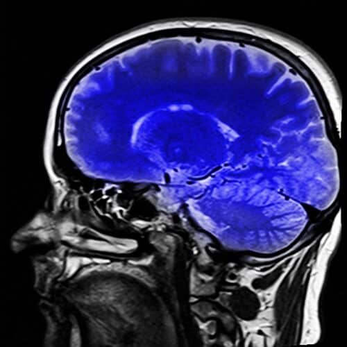
This new microscope succeeded in receiving a high-resolution image of the mouse brain’s neural network under the skull, without the need for removal of the skull and also without requiring a fluorescent label.
Click here for the Published Research Paper