

A revolutionary eye and brain implant design!
Researchers at the University of Oregon grew rodent retinal neurons on a fractal-patterned electrode that mimicked the repetitive branching pattern that occurs naturally in neurons. It brings physicist Richard Taylor of the University of Oregon one step closer to realising his long-held dream of creating a bio-inspired bionic eye.
Taylor hopes that the tiny electrodes will one day be put into the eye to help people with macular degeneration and other vision problems regain their vision. The latest research backs up a hunch his team has had for years: that neurons, which are fractals themselves, will attach better to a fractal-patterned electrode than to more conventionally shaped electrodes, allowing for improved signal transmission between the implant and the brain.
“The reason I’m so excited is that this paper is three years of data that explores what happens when these retinal cells interact with a fractal electrode,” he said. “You want neurons to get attached to be stimulated; that’s the ultimate goal in designing any sort of electrode,” said Saba Moslehi, a postdoctoral researcher in Taylor’s lab. “And when two objects have very similar characteristics, they’ll have more of a tendency to interact compared to objects that have completely different characteristics.”
The University of Oregon researchers employed electrodes fabricated from smooth silicon chips with carbon nanotube branches etched on the chip surface. Because neurons prefer to connect to textured nanotubes, researchers may manipulate the nanotube map on the electrode’s surface to determine where neurons attach. Moslehi and PhD students Conor Rowland and Julian Smith created silicon-based carbon nanotubes arrayed in a fractal pattern shaped like a repeating letter H (See the image given below) using facilities at the University of Oregon’s Center for Advanced Materials Characterization in Oregon.
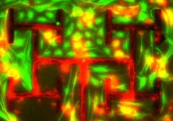
They also produced chips with the nanotubes arrayed in parallel lines, as seen on a commercially available electrode chip, for comparison. The researchers then used cells cultivated in a petri dish to track how mouse retinal neurons grew on the chips.
The experiment revealed that neurons connected more frequently to the textured fractal branches compared to the smooth intervals between the branches. Glia, which are crucial support cells for neurons, are crammed into the smooth spaces. This ‘herding’ of neurons and glia was most efficient with the fractal design.
Taylor stressed that the project is still in its early stages. However, the researchers hope that their concept can one day become a real-world device that can assist patients who have vision loss. Taylor and his colleagues published their findings in the journal PLOS One on April 6th 2022.