




The technology behind ultrasound imaging allows scientists to observe the development of eggs in living corals without resorting to any form of intrusive manipulation.
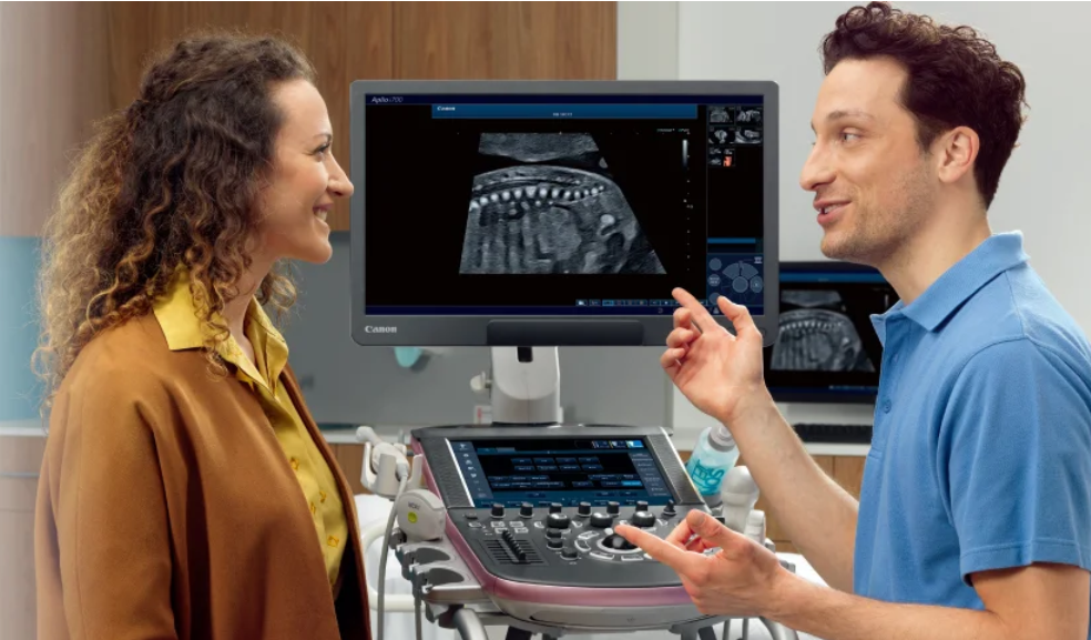
The medical technology of ultrasound allows the opening of new possibilities in the field of marine biology. Researchers can examine reproductive structures in live corals without cutting or injuring them. This method has been shown using Canon Medical Systems’ Aplio i700 ultrasound system.

The test was carried out by Coral Spawning International (CSI), along with Canon Medical Systems UK and Canon EMEA. Scientists used the ultrasound technology to scan six different LPS corals with various skeleton compositions while the corals were submerged under the water. The purpose of this procedure was to detect developing eggs inside the coral, which, until now, has entailed researchers cutting the corals to see their inner structure.
The use of high and ultra-high-frequency ultrasound probes on the Aplio i700 system was effective in generating images of the internal structure of corals. Ultrasound is a technique that involves the transmission of high frequency sound waves inside an object and analysis of the echoed waves coming back from the internal structures to form images.
In this research, ultrasound imaging technique offered a noninvasive approach to detecting the developing eggs without destroying the live corals. To verify the effectiveness of the imaging process, a specimen of Trachyphyllia geoffroyi was broken down and compared against the images obtained using macro photography.
This technique can be used in overcoming an important problem in conservation of corals. Knowing the stage of reproductive maturity will allow scientists to accurately predict spawning periods. These predictions are important in the context of planning coral propagation and restoration programs.
It is expected that CSI will continue observing scanned corals for six months and tracking the release of gametes as a means of comparison between reproductive development and actual spawning. In addition, the results of the study may give information on the reproductive cycles of different coral species.
This experiment demonstrates the possibility of using medical imaging technologies in applications other than medicine, for example, biological studies and analysis of live animals.