





A portable ultrasound device could help find breast cancer earlier, allow more frequent breast checks, and help doctors monitor patients at home or in clinics.
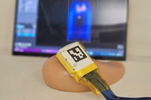
Researchers at the Massachusetts Institute of Technology (MIT) have developed a portable ultrasound system that could allow breast cancer screening to be performed more often, especially for people at high risk of developing the disease. The latest version produces clearer images and includes a guided interface that allows people without ultrasound training to perform scans.

The system is designed to detect tumors, cysts, and microcalcifications between annual mammograms. It can also be used to monitor patients after breast cancer treatment in a clinic or at home.
The research team improved image quality by adding a backing layer to the ultrasound transducer. The layer focuses sound waves, reduces acoustic and electrical noise, and supports a wider frequency range, resulting in clearer images.
The researchers also developed an adaptive beamforming algorithm that adjusts for differences in how sound waves travel through tissues such as skin and fat. This improves image reconstruction and increases image resolution by up to 10%.

To evaluate the system, 10 volunteers with no ultrasound experience were asked to locate embedded targets inside a breast tissue model. Participants identified the targets more often using the new system than with a conventional ultrasound probe.
The team also developed a computer interface that guides users to place the probe in the same position during each scan. This allows repeated scans from the same location, which is useful for monitoring treatment progress or tracking conditions such as fibroadenomas and microcalcifications.
In another trial involving seven participants, users were able to place the probe in the required position during repeated scans using the interface.
The system builds on an earlier prototype consisting of an ultrasound probe connected to a processing module about the size of a smartphone. It captures 3D images of the breast by scanning two or three locations.
The researchers plan to adapt the interface for smartphones and tablets, making the system easier to carry and use outside hospitals. They also believe it could increase access to ultrasound imaging in regions with limited availability of trained technicians.