

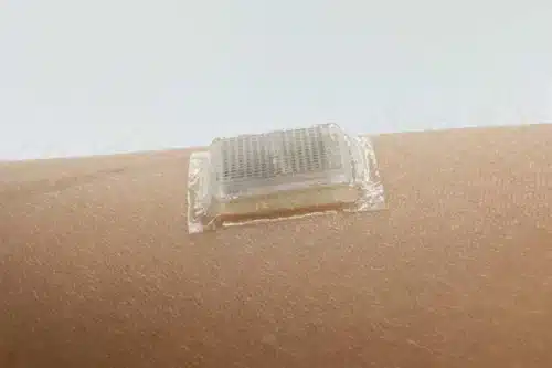
MIT Engineers invented an adhesive stamp-sized device that produces ultrasound images of the body.
The adhesive patch was developed to adhere to the body and produce continuous ultrasound images for 48 hours. The device needs to be connected to an instrument to convert reflected sound waves to images. The immediate goal of the researchers is to make this device wireless in such a way that patients can take home this wearable imaging device from a doctor or even buy it at a pharmacy
In ancient methods to record ultrasound images, technicians use to apply gel to patients’ skin that acts as a transmitter of sound waves. A probe or transducer is used to send sound waves into the body that reflect from the internal structures of the body back into the probe where echoed sound waves are translated into images. The disadvantage of this method for the patients who need a long time monitoring the probes had to affix for longer duration robotic arms were used to avoid long term holding in place but drying out of ultrasound liquid gel can interrupt the long-term imaging process. Recently, researchers have developed stretchable ultrasound probes which provide low-resolution images of internal organs but the stretchable property of probes may provide distorted images which may result in the inefficiency of imaging deep organs.’
The combination of a stretchy adhesive layer with a rigid array of transducers provides high-resolution images of internal organs. The exact location of transducers provides clearer and more precise images. The adhesive layer of the device is made up of two thin layers of elastomer that enclose a middle layer of solid hydrogel, which is a water-based material that easily transmits sound waves. The MIT team’s hydrogel is elastic and stretchy for high precision.
“With imaging, we might be able to capture the moment in a workout before overuse, and stop before muscles become sore,” says Chen, an MIT postdoc. “We do not know when that moment might be yet, but now we can provide imaging data that experts can interpret.”

The team is working to make the whole device wireless to make it more portable to use. The researchers of MIT envision Ultrasound stickers could be purchased by patients and consumers not only to image internal body organs but also to examine the progression of tumors or development of fetuses in the womb.
Click Here for a detailed video by MIT researchers.