


MRI can finally see and steer tiny medical robots at the same time. A scanning method removes delays and artifacts that once made real-time control impossible.
Real-time control of magnetic microrobots inside the body has been limited by MRI itself. Standard MRI scans are too slow, create artifacts, and interfere with the magnetic fields used to move the robots. This makes precise navigation difficult during live medical procedures, especially for clinicians and researchers working on minimally invasive interventions.
Researchers at Huazhong University of Science and Technology address this problem by redesigning the MRI scanning sequence. Their approach allows microrobots to be tracked in near real time without artifacts, while still using MRI to both image and drive the robot.
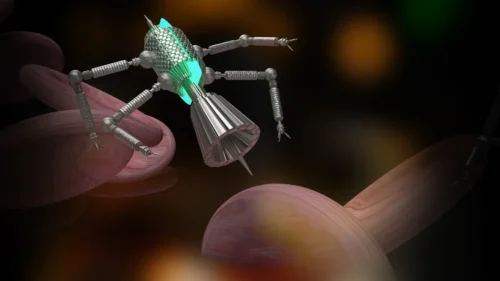
Magnetic microrobots are being explored for tasks such as targeted drug delivery, local therapy, and combined diagnosis and treatment. They can move through narrow and complex biological spaces that conventional tools cannot reach. MRI is attractive for guiding these robots because it images deep tissue with high spatial accuracy. The problem is speed. Conventional MRI repetition times are around 1,000 milliseconds, which introduces delays, reduces tracking accuracy, and disrupts the magnetic gradients needed for robot motion.
The new method uses a multi-frequency dual-echo MRI sequence that cuts repetition time to 30 milliseconds. This short cycle enables near real-time position updates. Two closely spaced radio-frequency pulses generate dual echoes to speed up signal recovery, while alternating frequency offsets prevent signal loss at high scan rates.
With this setup, robot position can be tracked with less than 1 percent relative error. Imaging and actuation no longer compete for gradient time, allowing a 77 percent driving duty cycle and clean background images. A custom reconstruction method replaces artifacts with bright markers on a pre-recorded background, so the robot remains visible while moving.
The system was tested in scenarios relevant to medical navigation. A robot was first guided through a complex 3D maze with live position updates. It then moved through vessel-like phantom models with tight curves, showing relevance for endovascular procedures. In an in vivo test, the robot navigated the large intestine of a rat under MRI guidance, pointing to potential use in gastrointestinal procedures where conventional tools are limited.
By removing MRI speed and interference limits, the technique makes MRI-guided microrobotics more practical for researchers and clinicians working on future minimally invasive therapies.