

A new microscope method reveals details that were difficult to see before, helping researchers study proteins, cells, and disease in new ways.
Researchers at Lawrence Berkeley National Laboratory and the University of California, Berkeley have developed a new technology that improves the performance of cryo-electron microscopes, enabling scientists to capture clearer images of small molecules and cellular structures that are difficult to study with existing systems.

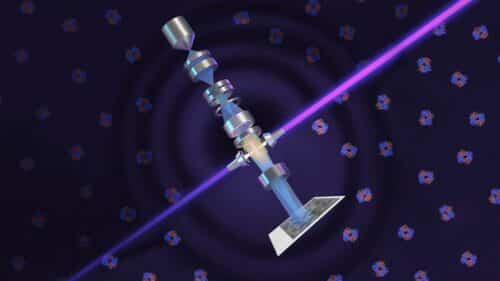
The team adapted phase-contrast imaging for cryo-electron microscopy (cryo-EM), a technique capable of magnifying samples about 10,000 times more than conventional light microscopes. The approach uses a laser-based phase plate that enhances image quality, allowing researchers to visualize biological structures that are often challenging for even the most advanced cryo-EM instruments.
The phase plate is integrated with a custom microscope developed in collaboration with Thermo Fisher Scientific. Designed to take advantage of the phase plate’s high-intensity laser, the system produces sharper images with greater detail. The improved image quality enables structure-analysis software to generate more accurate atomic models of the molecules being studied.
According to the research team, the technology is the result of more than 15 years of theoretical and experimental work involving microscopy researchers, machinists, and support from Biohub.

To demonstrate the system’s capabilities, the researchers imaged aldolase, a muscle protein that can already be analyzed using current cryo-EM systems, and hemoglobin, the oxygen-carrying protein in blood. Hemoglobin is significantly smaller and is commonly used as a benchmark for evaluating cryo-EM performance because it approaches the lower size limit that current instruments can reliably resolve.
The laser phase plate improved the structural resolution of both proteins, with the most significant gains observed for hemoglobin and other difficult samples. Researchers noted that the technology delivers the greatest benefits when studying small particles or specimens that are challenging to prepare and image.
The microscope, named Theia, is currently installed at UC Berkeley. The research team is now working to extend its capabilities beyond single-particle analysis to support cryo-electron tomography (cryo-ET), a technique that reconstructs three-dimensional images from multiple viewing angles.