

Scientists develop advanced techniques to observe the formation of platinum-nickel nanoparticles at atomic levels for material innovations.
Researchers from the Institute of Nuclear Physics, Polish Academy of Sciences, Poland have taken nanotechnology a step ahead by observing the real-time synthesis of platinum-nickel (PtNi) nanoparticles. Using advanced imaging techniques, they demonstrated the electrodeposition of nanostructured films on carbon electrodes, crucial for improving applications in energy and medicine.
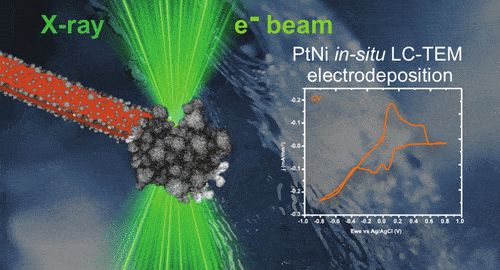
Electrodeposition, a widely used technique, involves immersing an electrode in a metal salt solution and applying voltage to grow nanoparticle layers. While this method is efficient, its complexities at the atomic scale remained elusive until now. The team used liquid-cell transmission electron microscopy (TEM) to monitor these processes directly, overcoming traditional imaging limitations of sample thickness and dryness.
“By using a liquid flow cell in TEM, we could observe nucleation and nanoparticle growth in real time,” said Magdalena Parlińska-Wojtan, professor, IFJ PAN. Their setup employed electron-transparent silicon membranes, creating precise imaging of the nanoparticles’ development. This development holds immense potential for industries like renewable energy and advanced medical therapies, where the demand for highly efficient, precisely designed materials continues to rise.
Experiments revealed that PtNi films grow in two ways: nanoparticles nucleate on the electrode or form in the surrounding solution before attaching. Subsequent dry observations showed that the films consist of branched, spherical nanoparticles—a structure critical for their catalytic efficiency.
In collaboration with the Fritz Haber Institute, Berlin, researchers modified electrodeposition conditions to study the nanoparticles’ growth dynamics. They observed alternating growth and dissolution during voltage cycles, with growth dominating, leading to stable nanolayers.
The team also used scanning transmission X-ray microscopy (STXM) at Kraków’s SOLARIS center to investigate material properties. STXM revealed that the resulting layers comprise both metallic platinum and nickel oxide, demonstrating the method’s versatility for studying oxidation states.
Parlińska-Wojtan emphasised the significance of this research, stating, “Understanding the synthesis and growth of nanostructures will enable tailored designs for fuel cells, medical applications, and beyond.”
The study highlights the potential of imaging to drive innovation in nanotechnology.